

The article titled "10 Key Insights on PET Myocardial Perfusion Imaging for Researchers" emphasizes the advancements, applications, and clinical significance of PET myocardial perfusion imaging (MPI) in cardiac diagnostics. It underscores that PET MPI is essential for accurately diagnosing coronary artery disease (CAD) and assessing myocardial viability. This is supported by its superior sensitivity and specificity compared to other imaging modalities, such as SPECT. Moreover, advancements in technology and the integration of AI enhance its diagnostic capabilities, making it an invaluable tool in the Medtech landscape.
The realm of cardiac diagnostics is rapidly evolving, with PET myocardial perfusion imaging emerging as a pivotal tool in the assessment of heart health. This advanced imaging technique not only offers unparalleled insights into myocardial blood flow but also significantly enhances diagnostic accuracy in detecting coronary artery disease. As researchers delve into the intricacies of PET imaging, a pressing question arises: how can the latest advancements and strategic partnerships reshape the future of cardiac care? Exploring key insights into PET myocardial perfusion imaging reveals a landscape rich with potential, poised to transform clinical practices and improve patient outcomes.
bioaccess® excels in accelerating clinical research for pet myocardial perfusion imaging by leveraging the regulatory efficiency of Latin America, the diverse demographics of the Balkans, and the streamlined ethical approval processes in Australia. This strategic combination ensures ethical approvals are achieved within 4-6 weeks, significantly enhancing patient enrollment rates by 50% compared to traditional markets. As a vital partner for Medtech, Biopharma, and Radiopharma innovators, bioaccess® enables clients to expedite their product development timelines through comprehensive services, including:
Recent advancements in clinical research underscore the critical need for swift trial initiation, with the global clinical trial visual assessment market projected to reach USD 1.91 billion by 2030, expanding at a CAGR of 7.8%. Furthermore, successful partnerships in clinical research are increasingly essential; 57% of industry professionals identify regulatory hurdles as a primary cause of delays in product launches. By leveraging its expertise and innovative strategies, including collaboration with the Caribbean Health Group and support from Colombia's Minister of Health, bioaccess® not only accelerates ethical approvals but also empowers its clients to seize new opportunities in the rapidly evolving field of pet myocardial perfusion imaging.

Positron Emission Tomography (PET) scans operate on the principle of detecting gamma rays emitted from a radioactive tracer introduced into the bloodstream. In the assessment of heart blood flow, tracers such as Rubidium-82 or Nitrogen-13 ammonia are commonly employed. These tracers facilitate PET myocardial perfusion imaging, which allows for the visualization of blood circulation to the heart tissue and provides critical insights into heart muscle viability and perfusion across various physiological conditions.

Effective assessment of pet myocardial perfusion imaging is paramount in clinical settings, necessitating strict adherence to established protocols. This includes essential patient preparation strategies such as:
The American Society of Nuclear Cardiology (ASNC) provides comprehensive guidelines advocating for a rest-stress protocol that incorporates pet myocardial perfusion imaging to accurately evaluate coronary blood flow by capturing images both at rest and during pharmacological stress. Furthermore, the proper calibration of the PET scanner, alongside rigorous quality control measures, is crucial to ensure the production of high-quality visual results.

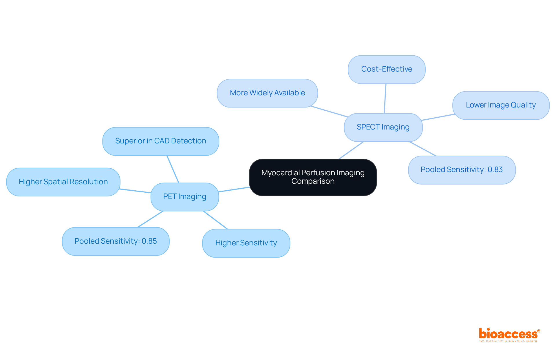
Both Positron Emission Tomography (PET) myocardial perfusion imaging and Single Photon Emission Computed Tomography (SPECT) are employed for heart perfusion evaluation; however, PET myocardial perfusion imaging distinctly surpasses SPECT in several critical aspects. Notably, PET offers higher spatial resolution and sensitivity, enabling the detection of smaller quantities of radioactivity, which is essential for precise imaging. This enhanced sensitivity proves particularly advantageous in clinical settings, where accurate measurement of absolute heart blood flow is vital for diagnosing conditions such as coronary artery disease (CAD).
Recent studies consistently demonstrate that PET myocardial perfusion imaging is superior in identifying CAD and assessing myocardial viability. For instance, a meta-analysis encompassing 12 studies with a total sample size of 397 participants revealed that PET scans yield a pooled sensitivity of 0.85, compared to SPECT's pooled sensitivity of 0.83, which ranged from 59% to 95%. Furthermore, PET's diagnostic accuracy is strengthened by its ability to visualize low quantities of radioactivity, rendering it invaluable in early-phase drug development and clinical research.
While SPECT remains more widely available and cost-effective, its lower image quality and diagnostic accuracy can restrict its effectiveness in high-stakes clinical scenarios. As Timothy M. Bateman notes, "Emerging evidence consistently demonstrates that PET provides improved image quality, greater interpretive certainty, and higher diagnostic accuracy compared to SPECT." This positions PET as the preferred choice for many clinicians, particularly when evaluating patients with an intermediate likelihood of significant CAD. Additionally, it is crucial to acknowledge that approximately 20% of the yearly total radiation dose received by the United States population from diagnostic procedures arises from radionuclide MPI, underscoring the importance of selecting techniques that minimize radiation exposure. Overall, the advantages of PET myocardial perfusion imaging in heart perfusion assessment highlight its essential role in advancing cardiac diagnostics.

Extensive research has established that pet myocardial perfusion imaging (MPI) using Rb-82 offers exceptional diagnostic accuracy for detecting obstructive coronary artery disease (CAD). Studies indicate that Rb-82 PET achieves sensitivity rates of 90% (CI: 0.88 to 0.92) and specificity rates around 88% (CI: 0.85 to 0.91), significantly surpassing SPECT in various clinical scenarios.
The capability to measure myocardial blood flow (MBF) and evaluate myocardial flow reserve (MFR) enhances pet myocardial perfusion imaging's diagnostic abilities, facilitating better risk stratification and management for individuals with suspected CAD. Notably, patients with normal pet myocardial perfusion imaging scans demonstrate a 0% annualized event rate for hard cardiac events, which underscores the prognostic value of pet myocardial perfusion imaging.
Furthermore, the integration of coronary artery calcium scoring with pet myocardial perfusion imaging has been demonstrated to enhance diagnostic and prognostic accuracy, making it an essential tool in contemporary cardiovascular assessment.
As specialists emphasize, "The capability to diagnose subclinical CAD is a robust characteristic of PET technology," highlighting its benefits in clinical practice.

Pet myocardial perfusion imaging serves a dual purpose: diagnosing coronary artery disease (CAD) and providing critical prognostic insights. Recent research indicates that individuals exhibiting decreased heart muscle blood flow or compromised heart muscle flow reserve face a significantly heightened risk of major adverse cardiac events (MACEs), including heart attacks and mortality. Notably, an increase in stress myocardial blood flow (MBF) by just 1 mL/g/min correlates with a protective hazard ratio of 0.32 for MACEs, while high-stress MBF is linked to a hazard ratio of 0.43, emphasizing the vital role of MBF quantification in risk assessment.
Furthermore, the annualized event rate (AER) for diabetics stands at 1.4%, compared to 0.3% for nondiabetics, highlighting the increased risk for those with diabetes. By incorporating PET scan findings into clinical decision-making, healthcare providers can enhance risk stratification and customize treatment strategies, ultimately leading to improved outcomes for patients. This integration of diagnostic data not only aids in identifying high-risk individuals but also facilitates timely interventions, thereby improving the management of CAD and its associated complications. However, further validation through large prospective randomized controlled trials is essential to solidify these findings.

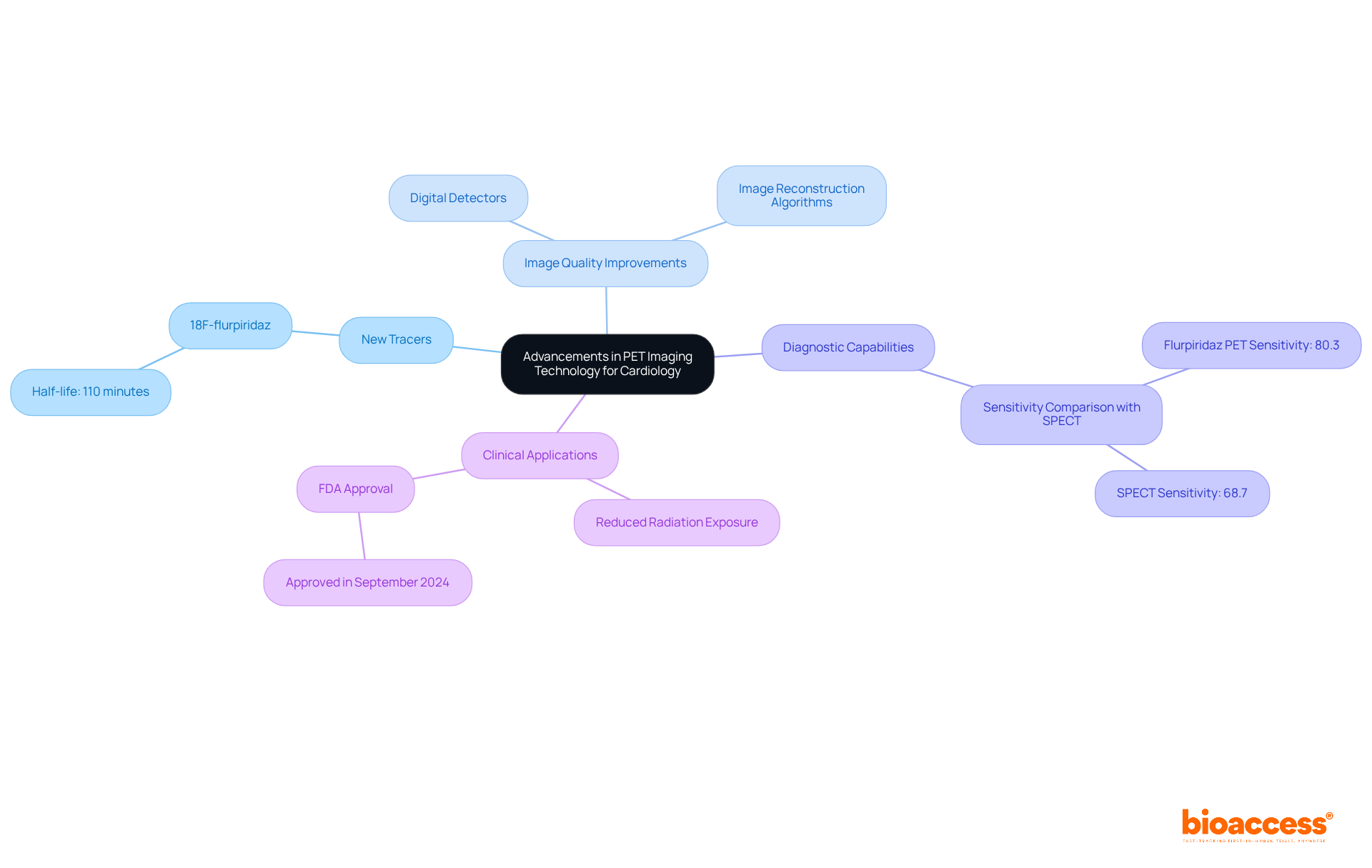
Recent advancements in PET myocardial perfusion imaging technology, particularly the introduction of new tracers such as 18F-flurpiridaz, have significantly enhanced the precision and effectiveness of heart perfusion assessment (MPI). Flurpiridaz, characterized by its extended half-life of approximately 110 minutes, facilitates more adaptable protocols and improved visualization of myocardial perfusion.
Research indicates that flurpiridaz PET exhibits superior diagnostic capability, with a sensitivity of 80.3% compared to 68.7% for conventional SPECT. Furthermore, advancements in PET scanner technology, including digital detectors and refined image reconstruction algorithms, have markedly improved image quality, resulting in a greater proportion of PET scans rated as good to excellent relative to SPECT.
These technological improvements also contribute to reduced radiation exposure for patients, establishing PET myocardial perfusion imaging as a more viable and effective option in clinical practice, particularly for the assessment of coronary artery disease (CAD). Notably, flurpiridaz F-18 received FDA approval in September 2024, representing a significant milestone in its clinical application.

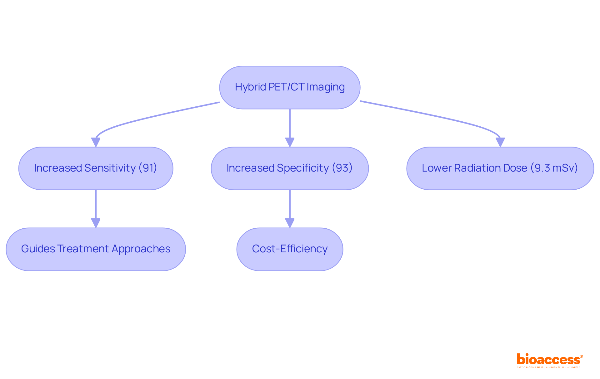
Hybrid PET/CT scans effectively combine the functional insights of PET with the anatomical precision of CT, delivering a comprehensive assessment of pet myocardial perfusion imaging. This integration not only facilitates the accurate localization of perfusion defects but also enhances the characterization of coronary artery disease (CAD).
Notably, studies reveal that hybrid techniques achieve a pooled sensitivity of 91% and specificity of 93% for detecting obstructive CAD, significantly surpassing standalone modalities. The negative likelihood ratio (LR−) of 0.11 for hybrid techniques, compared to 0.06 for CCTA, further underscores its diagnostic superiority.
The ability to visualize both functional and structural abnormalities enhances diagnostic precision and guides treatment approaches, establishing hybrid techniques as an essential resource in modern cardiology. Experts advocate for its regular application, with P. Knaapen emphasizing that this diagnostic technique is often referred to as the 'one-stop-shop' for cardiac evaluation.
Additionally, the hybrid approach presents a significant radiation dose advantage, averaging 9.3 mSv, which is lower than traditional methods. As research continues to validate its efficacy, pet myocardial perfusion imaging using hybrid PET/CT technology is poised to redefine standards in cardiac assessment, offering a comprehensive approach that aligns with the evolving landscape of cardiovascular diagnostics.
Moreover, the economic implications of hybrid techniques suggest potential reductions in subsequent testing and unnecessary procedures, marking it as a cost-efficient strategy for managing patients. While the benefits are substantial, ongoing studies are essential to explore its limitations and future directions within clinical practice.

PET myocardial perfusion imaging is an invaluable tool in various clinical scenarios, especially for assessing unusual presentations, evaluating myocardial viability, guiding revascularization decisions, and monitoring treatment response.
These scenarios underscore the versatility and clinical importance of PET scans in managing cardiac health, particularly in complex cases where traditional methods may not provide sufficient clarity. The American Society of Nuclear Medicine endorses PET myocardial perfusion imaging as a preferred diagnostic method, highlighting its diagnostic superiority and reduced radiation exposure compared to conventional techniques. This positions PET myocardial perfusion imaging as a crucial tool in the evolving landscape of cardiac diagnostics.

The future of pet myocardial perfusion imaging in cardiology is exceptionally promising, driven by ongoing research aimed at enhancing pet myocardial perfusion imaging techniques, developing innovative tracers, and integrating artificial intelligence (AI) for superior image analysis. Recent studies suggest that the integration of AI in pet myocardial perfusion imaging has the potential to significantly enhance diagnostic accuracy and efficiency, as AI algorithms demonstrate improved predictive abilities in detecting coronary artery disease (CAD) and evaluating myocardial perfusion irregularities.
As we approach 2025, research trends highlight a growing focus on the development of novel PET tracers that offer improved sensitivity and specificity for detecting CAD. For instance, Flurpiridaz PET has shown a sensitivity of 80.3% compared to 68.7% for SPECT, and a recent meta-analysis indicated a sensitivity of 91% and specificity of 86% for pet myocardial perfusion imaging studies. These advancements are complemented by the growth of hybrid visualization technologies, such as the D-SPECT system, which offers high-speed scanning for pet myocardial perfusion imaging with enhanced sensitivity compared to traditional SPECT cameras, allowing for more thorough assessments of cardiac health.
Furthermore, the incorporation of AI is anticipated to transform the evaluation of PET scan data, enabling more accurate risk categorization and tailored treatment approaches for individuals with cardiovascular conditions. This evolution positions pet myocardial perfusion imaging as a cornerstone in cardiac diagnostics, enhancing its role in personalized medicine and improving patient outcomes in managing cardiovascular conditions. However, challenges such as the need for specialized workflows and higher costs may hinder widespread adoption, necessitating ongoing economic analyses to assess the cost-effectiveness of these advancements.

The insights gathered on PET myocardial perfusion imaging highlight its transformative role in cardiac diagnostics, particularly as advancements in technology and research continue to unfold. This imaging modality not only enhances the accuracy of diagnosing coronary artery disease but also provides critical prognostic information that can significantly influence patient management and outcomes.
Key arguments presented emphasize the advantages of PET over traditional SPECT imaging, including:
Furthermore, the integration of innovative tracers and hybrid imaging technologies is poised to elevate the standard of care in cardiology, rendering PET an invaluable tool in the clinical setting.
As the landscape of cardiac imaging evolves, it is essential for researchers, clinicians, and stakeholders to embrace these advancements and continue exploring the potential of PET myocardial perfusion imaging. By prioritizing further research and collaboration, the medical community can enhance diagnostic capabilities, ultimately leading to improved patient care and outcomes in cardiovascular health.
What is bioaccess® and how does it support clinical research for PET myocardial perfusion imaging?
bioaccess® accelerates clinical research for PET myocardial perfusion imaging by utilizing the regulatory efficiency of Latin America, diverse demographics of the Balkans, and streamlined ethical approval processes in Australia. This combination allows for ethical approvals within 4-6 weeks and enhances patient enrollment rates by 50% compared to traditional markets.
What services does bioaccess® provide to its clients?
bioaccess® offers comprehensive services including feasibility studies, selection of research sites and principal investigators (PIs), and detailed reporting on study status, inventory, and adverse events.
What is the significance of advancements in clinical research for PET myocardial perfusion imaging?
Recent advancements highlight the need for swift trial initiation, with the global clinical trial visual assessment market expected to reach USD 1.91 billion by 2030, growing at a CAGR of 7.8%. Successful partnerships are also crucial, as 57% of industry professionals cite regulatory hurdles as a primary cause of product launch delays.
How does bioaccess® leverage partnerships to enhance its services?
bioaccess® collaborates with entities like the Caribbean Health Group and receives support from Colombia's Minister of Health to accelerate ethical approvals and empower clients to capitalize on new opportunities in PET myocardial perfusion imaging.
What is the principle behind PET imaging in myocardial perfusion?
PET imaging operates by detecting gamma rays emitted from a radioactive tracer introduced into the bloodstream, allowing for visualization of blood circulation to the heart tissue and providing insights into heart muscle viability and perfusion under various conditions.
What are the essential protocols for effective PET myocardial perfusion imaging?
Effective assessment requires strict adherence to protocols that include patient preparation strategies such as fasting, avoiding caffeine, careful tracer selection, and precise timing of scans.
What guidelines does the American Society of Nuclear Cardiology (ASNC) provide for PET myocardial perfusion imaging?
The ASNC advocates for a rest-stress protocol that incorporates PET myocardial perfusion imaging to accurately evaluate coronary blood flow by capturing images both at rest and during pharmacological stress.
Why is proper calibration and quality control important in PET imaging?
Proper calibration of the PET scanner and rigorous quality control measures are crucial to ensure the production of high-quality visual results in PET myocardial perfusion imaging.