

The article elucidates the critical necessity of comprehensive treatment for fixed perfusion defects (FPDs), given their association with severe conditions such as coronary artery disease and myocardial infarction, which considerably elevate mortality risks. It provides an in-depth examination of diagnostic techniques and treatment strategies, including medical therapy and revascularization. Furthermore, it underscores the vital role of long-term management and follow-up care in enhancing patient outcomes, thereby reinforcing the imperative of addressing these health challenges effectively.
Understanding fixed perfusion defects (FPDs) is crucial for effective cardiovascular care, as these conditions often signal underlying coronary artery disease and can significantly impact patient outcomes. This article delves into comprehensive strategies for diagnosing and treating FPDs, emphasizing the importance of early detection and tailored interventions.
How can healthcare professionals optimize treatment plans for patients with these defects?
What innovative approaches are emerging in the field of cardiology to enhance long-term management?
These questions are pivotal as we explore the landscape of cardiovascular health.
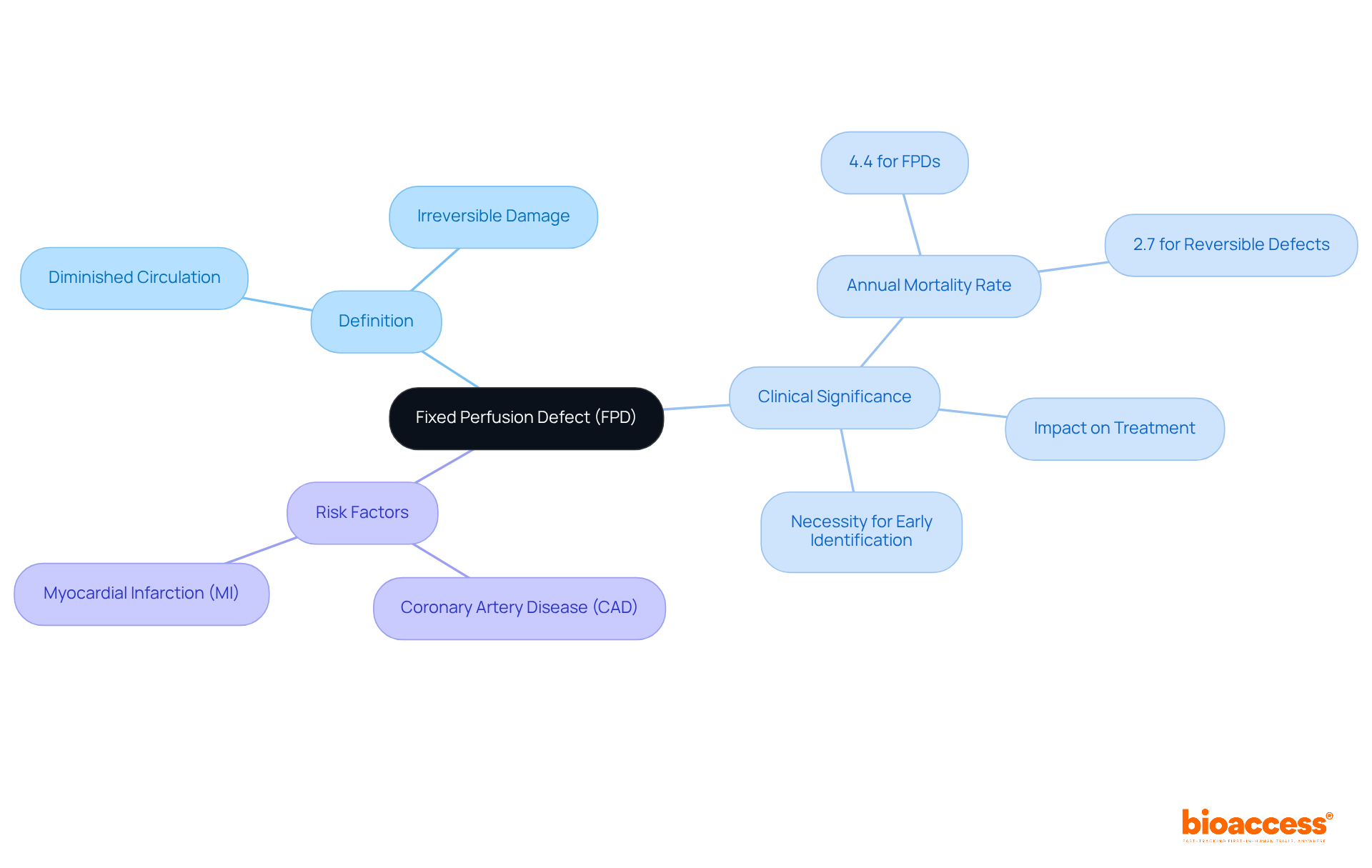
A fixed blood flow defect (FPD) represents an area of the heart muscle with diminished circulation observable during both rest and stress imaging, indicating irreversible damage or scarring. Clinically, FPDs are pivotal as they frequently denote underlying coronary artery disease (CAD) or a history of myocardial infarction (MI). Notably, recent studies reveal that individuals with FPDs face an annual mortality rate of 4.4%, significantly higher than the 2.7% observed in those with reversible perfusion abnormalities. This stark contrast accentuates the necessity for early identification of FPDs, which is essential for effective risk stratification and treatment for fixed perfusion defect.
Cardiologists assert that recognizing these defects is crucial for directing further diagnostic testing and the treatment for fixed perfusion defect, given that FPDs correlate with adverse outcomes, including heightened morbidity and mortality. For instance, a study involving 327 individuals without known CAD indicated that the presence of FPDs was linked to a risk ratio of 2.5 for increased mortality, underscoring their prognostic significance. Furthermore, it is noteworthy that only 5% of the examined group exhibited considerable left ventricular ischemia, providing additional context regarding the cardiac health of individuals with FPDs.
By comprehending the implications of FPDs, clinicians can make informed decisions that enhance patient outcomes in managing CAD.

Several diagnostic techniques are employed to identify fixed perfusion defects, including:
Myocardial Perfusion Imaging (MPI): This non-invasive imaging technique utilizes radioactive tracers to visualize blood flow to the heart muscle during both rest and stress conditions. Common modalities used in MPI include SPECT (Single Photon Emission Computed Tomography) and PET (Positron Emission Tomography).
Cardiac MRI: This imaging method provides intricate images of the heart's structure and function, enabling a thorough evaluation of myocardial blood flow and viability.
Angiography: Though invasive, this definitive technique visualizes the heart's arteries, effectively identifying blockages that may lead to fixed perfusion defects.
Computed Tomography Angiography (CTA): As a non-invasive imaging technique, CTA evaluates heart artery disease and is frequently utilized alongside MPI to enhance diagnostic precision.
Each of these techniques possesses distinct strengths and limitations. The choice of method often depends on the clinical situation and individual patient characteristics.

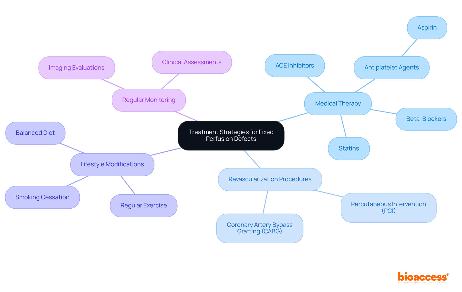
Treatment for fixed perfusion defect focuses on effectively addressing coronary artery disease (CAD) and enhancing outcomes for individuals. Key approaches include:
Medical Therapy: This typically involves antiplatelet agents such as aspirin, beta-blockers, ACE inhibitors, and statins, which are essential for managing cardiovascular risk factors and improving heart function. Recent guidelines emphasize the role of statins in lowering low-density lipoprotein (LDL) cholesterol, significantly reducing the risk of subsequent heart attacks after two years of treatment. As William C. Roberts, MD, noted, "Statin drugs, in my view, are the best cardiovascular drugs ever created."
Revascularization Procedures: For individuals with significant artery blockages, procedures like percutaneous intervention (PCI) or artery bypass grafting (CABG) are often necessary to restore blood flow. Studies indicate that CABG is associated with improved long-term survival and lower rates of major adverse cardiac and cerebrovascular events (MACCE) compared to PCI, particularly in patients with complex coronary anatomy. The FREEDOM trial found that CABG favored all-cause death and non-fatal myocardial infarction at five years.
Lifestyle Modifications: Encouraging heart-healthy lifestyle changes—such as a balanced diet, regular exercise, and smoking cessation—is crucial for long-term management. Evidence indicates that even moderate physical activity can greatly lower cardiovascular risks, making lifestyle interventions an essential part of care.
Regular Monitoring: Ongoing follow-up through imaging and clinical evaluations is essential to assess effectiveness and make necessary adjustments. This proactive approach helps in identifying any progression of CAD and tailoring interventions accordingly.
The choice of care should be customized, considering the individual's overall health, the severity of CAD, and any existing comorbidities, particularly in relation to the treatment for fixed perfusion defect. As cardiologists note, while revascularization can provide immediate relief, optimal medical therapy remains a cornerstone in managing CAD effectively.

Long-term management of patients with fixed perfusion defects necessitates a comprehensive approach that encompasses several critical strategies:
Regular Follow-Up Appointments: Scheduled visits are essential for monitoring cardiovascular health, evaluating the effectiveness of care, and adjusting medications as necessary.
Repeat Imaging: Periodic myocardial perfusion imaging is crucial to assess changes in perfusion status and inform further care decisions.
Client Education: Educating individuals about their condition, treatment alternatives, and the importance of adhering to prescribed therapies is vital for effective long-term management.
Multidisciplinary Care: Collaboration among cardiologists, primary care physicians, dietitians, and other healthcare professionals can significantly enhance health outcomes by addressing all aspects of well-being.
Psychosocial Support: Providing resources for mental health assistance is essential in helping individuals manage the emotional challenges associated with chronic conditions.
By implementing these strategies, healthcare providers can markedly improve the quality of life and prognosis for patients requiring treatment for fixed perfusion defect.

Understanding and managing fixed perfusion defects (FPDs) is crucial for improving patient outcomes in cardiovascular health. These defects serve as indicators of underlying coronary artery disease and can significantly impact mortality rates. Early identification and appropriate treatment strategies are essential, as they lead to better risk stratification and enhanced care for affected individuals.
The article delves into various aspects of FPDs, including their clinical significance, diagnostic techniques, treatment strategies, and long-term management. Recognizing FPDs is important to direct further testing and treatment. A range of diagnostic tools is available, such as:
Additionally, multifaceted treatment approaches encompass:
The necessity of a comprehensive follow-up care plan is emphasized, including client education and multidisciplinary collaboration to ensure optimal health outcomes.
Ultimately, addressing fixed perfusion defects is not just about immediate treatment; it is about fostering a proactive approach to long-term management. By prioritizing early detection, personalized treatment plans, and ongoing support, healthcare providers can significantly enhance the quality of life for individuals with FPDs. This commitment to comprehensive care underscores the importance of staying informed about the latest advancements in fixed perfusion defect management and the critical role that proactive health strategies play in improving patient outcomes.
What is a fixed perfusion defect (FPD)?
A fixed perfusion defect (FPD) is an area of the heart muscle that shows diminished blood flow during both rest and stress imaging, indicating irreversible damage or scarring.
Why are fixed perfusion defects clinically significant?
FPDs are significant because they often indicate underlying coronary artery disease (CAD) or a history of myocardial infarction (MI). They are associated with higher morbidity and mortality rates.
What is the annual mortality rate for individuals with fixed perfusion defects?
Individuals with fixed perfusion defects face an annual mortality rate of 4.4%, which is significantly higher than the 2.7% observed in those with reversible perfusion abnormalities.
How do fixed perfusion defects affect patient management?
Recognizing FPDs is crucial for directing further diagnostic testing and treatment, as they correlate with adverse outcomes, including increased morbidity and mortality.
What does research indicate about the prognostic significance of fixed perfusion defects?
A study found that the presence of FPDs was linked to a risk ratio of 2.5 for increased mortality in individuals without known CAD, highlighting their prognostic importance.
What percentage of individuals with fixed perfusion defects exhibited considerable left ventricular ischemia?
Only 5% of the examined group with fixed perfusion defects showed considerable left ventricular ischemia, providing context regarding their cardiac health.
How can understanding fixed perfusion defects improve patient outcomes?
By understanding the implications of FPDs, clinicians can make informed decisions that enhance patient outcomes in managing coronary artery disease.