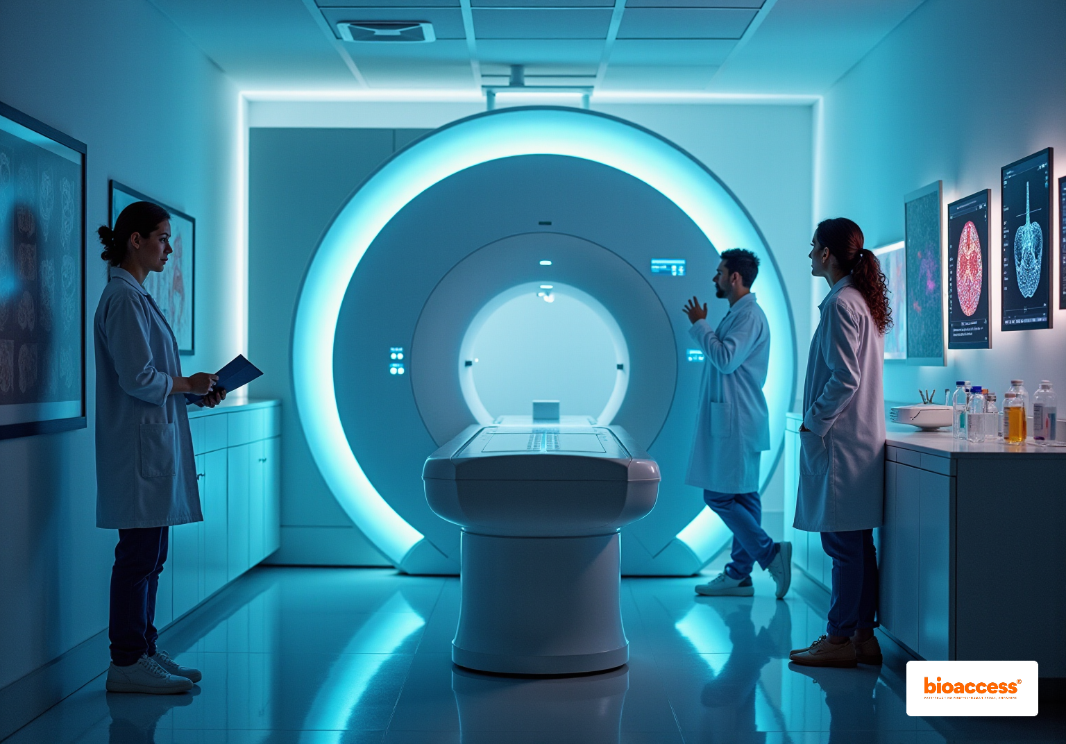


Tracer imaging stands as a cornerstone technique in clinical research, utilizing radioactive markers to visualize and measure biological processes. This method significantly enhances our understanding of disease mechanisms and treatment efficacy.
With advancements in imaging technologies such as PET and SPECT, diagnostic accuracy has improved, leading to better patient outcomes. These developments showcase the technique's critical role in early diagnosis and personalized medicine, making it an essential tool for researchers and clinicians alike.
Tracer imaging stands at the forefront of clinical research, providing critical insights into the complex workings of biological processes through radioactive markers. This powerful technique not only enhances our understanding of drug distribution and disease progression but also plays a pivotal role in improving patient outcomes. By facilitating early diagnosis and tailored treatment strategies, tracer imaging is essential in the modern medical landscape.
As advancements in imaging technology continue to emerge, clinical researchers face the challenge of effectively implementing and interpreting these complex methodologies. How can they navigate this evolving landscape to maximize the benefits of tracer imaging while overcoming potential hurdles? Addressing these questions is vital for harnessing the full potential of this innovative approach in clinical settings.
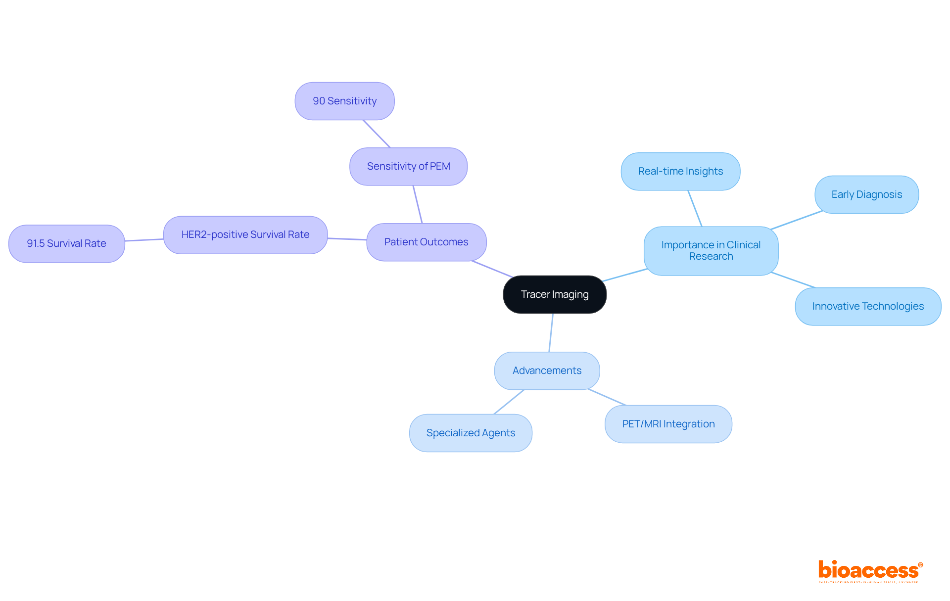
Tracer imaging techniques utilize radioactive materials, known as markers, to observe and measure biological processes within the body. This approach is crucial in clinical research, enabling the observation of drug distribution, monitoring disease progression, and assessing treatment efficacy. By offering real-time insights into physiological functions, scanning deepens our understanding of complex biological systems, ultimately enhancing patient outcomes. Its importance is highlighted by its ability to facilitate early diagnosis, inform therapeutic decisions, and propel the development of innovative medical technologies.
Recent advancements in visualization methods, particularly the integration of PET with MRI, have shown remarkable improvements in diagnostic accuracy. For example, studies reveal that this combination can boost the positive predictive value from 77% to an impressive 98%, significantly improving the detection of malignant lesions. Additionally, the use of specialized agents like Ga-FAPI has demonstrated superior tumor absorption and metastatic detection rates compared to traditional methods, especially in challenging cases such as invasive lobular carcinoma.
Data underscore the substantial impact of tracer imaging techniques on patient outcomes. For instance, the five-year survival rate for individuals with HER2-positive breast cancer can soar to 91.5% when effectively monitored and treated using advanced scanning methods. Moreover, the sensitivity of positron emission mammography (PEM) in identifying malignant lesions has been reported at 96%, showcasing its potential in preoperative staging and treatment planning. The five-year relative survival rates for breast cancer subtypes further illustrate this effect, with luminal A at 94.4% and luminal B at 90.7%.
As tracer imaging continues to evolve, its role in medical research becomes increasingly vital, paving the way for more personalized and efficient patient care.
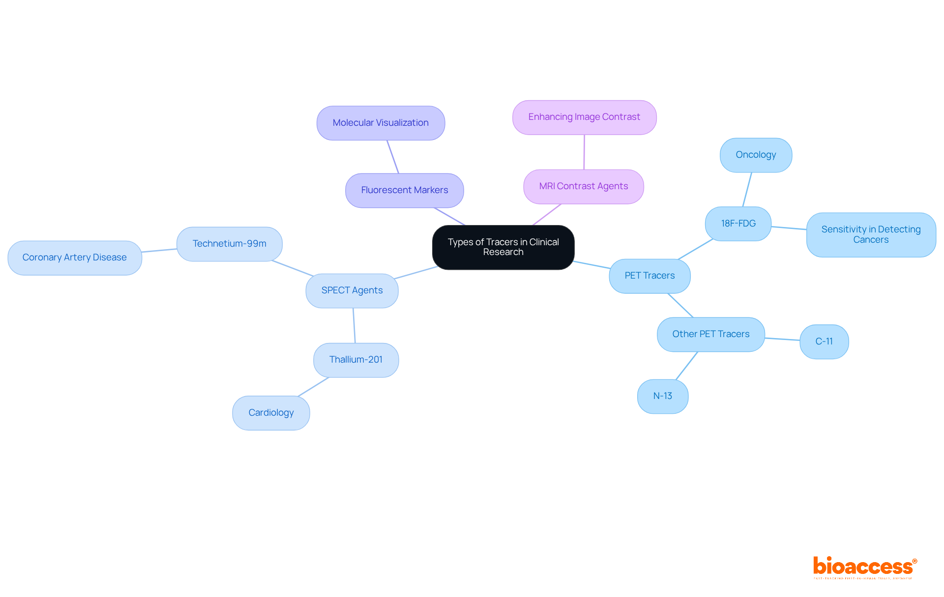
In clinical research, tracer imaging and various types of tracers are employed, each with specific applications that significantly impact patient management and treatment outcomes.
Positron Emission Tomography (PET) Tracers: Predominantly utilized in oncology, PET tracers such as 18F-fluorodeoxyglucose (FDG) are essential for visualizing metabolic activity in tumors. PET imaging has demonstrated a sensitivity of approximately 95% in detecting esophageal carcinoma and an accuracy of 96% for staging lymphomas. This capability significantly aids in diagnosis and staging. Furthermore, FDG can identify primary colon carcinoma in over 90% of instances, compared to the 60% sensitivity of CT, highlighting the efficacy of PET agents in oncology.
Single Photon Emission Computed Tomography (SPECT) Agents: SPECT agents are primarily used in cardiology to evaluate blood flow and heart function. For instance, myocardial perfusion studies utilize substances such as thallium-201 and technetium-99m to assess coronary artery disease, providing essential information about cardiac health. Additionally, MPI-PET offers valuable insights into coronary flow reserve, aiding in the evaluation of individual coronary artery lesions.
Fluorescent Markers: These markers are crucial for molecular visualization, enabling researchers to observe cellular processes at the molecular level. Their application is vital in understanding disease mechanisms and developing targeted therapies.
MRI Contrast Agents: Enhancing the contrast of images in magnetic resonance imaging, these agents facilitate the diagnosis of various conditions by improving the visibility of internal structures.
Each type of marker possesses unique properties tailored to specific research objectives, making it essential for researchers to choose the suitable marker based on the biological processes they aim to investigate. The integration of PET and SPECT imaging agents in clinical settings has transformed oncology and cardiology, offering real-time insights through tracer imaging that greatly influence patient management and treatment results.
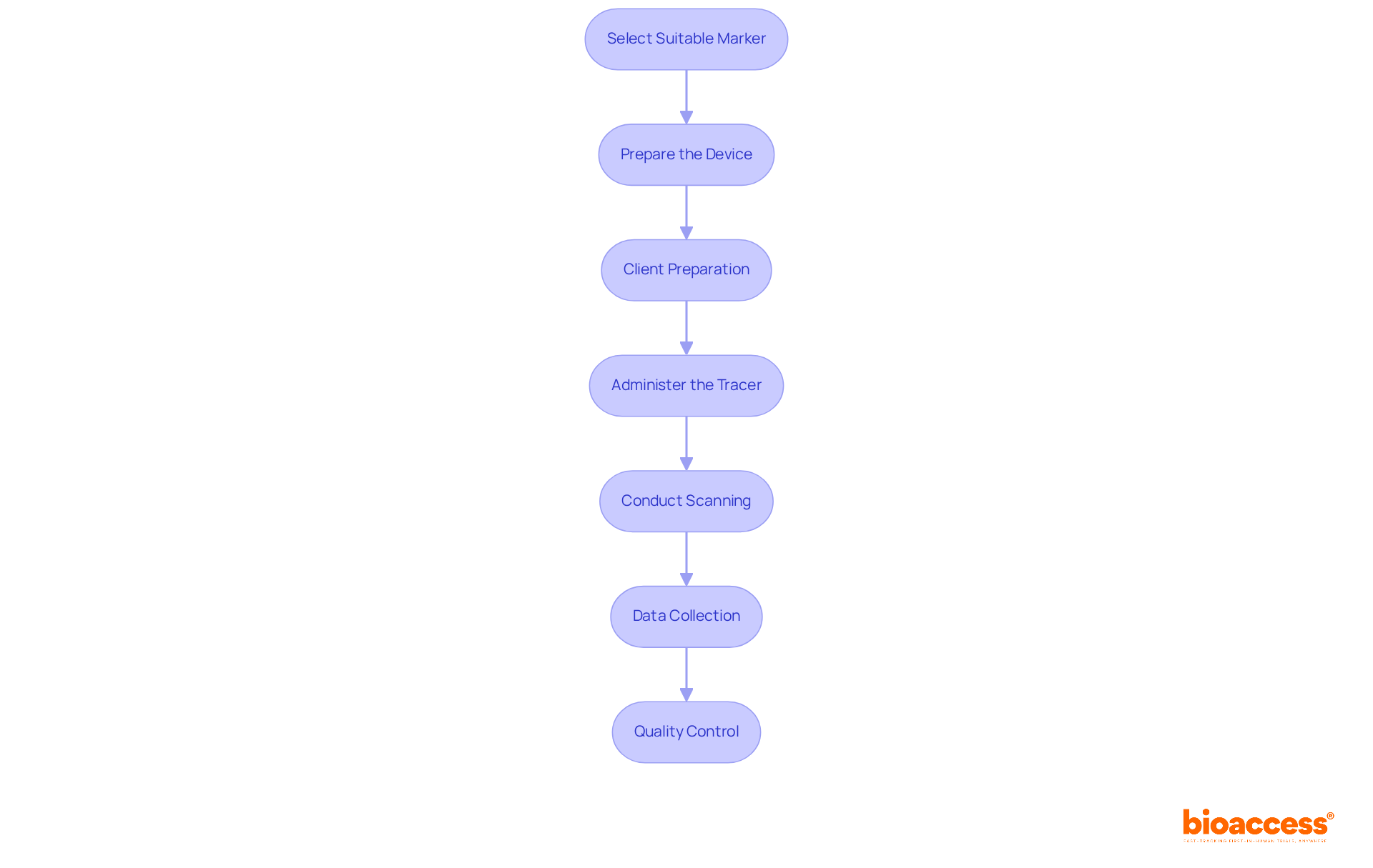
To implement tracer imaging effectively in clinical research, it’s essential to follow these best practices:
Select the Suitable Marker: Choose a marker that aligns with the biological process under investigation, considering the specific disease context. For instance, individuals with malignant diseases are typically more appropriate for radioactive ligands, while benign conditions may justify their application if they significantly affect morbidity or prognosis.
Prepare the Device: Ensure the device is prepared in compliance with safety and regulatory guidelines, preserving its integrity for accurate results.
Client Preparation: Clearly inform individuals about the procedure, emphasizing its purpose and any potential risks. This step is crucial, as medical ethical committees evaluate the use of radionuclides based on the disease's morbidity and prognosis.
Administer the Tracer: Administer the tracer via the appropriate route, such as intravenous or oral, while closely monitoring the patient for any adverse reactions. The timing of administration should consider the half-life of the therapeutic compound to enhance diagnostic results.
Conduct Scanning: Utilize relevant visualization technologies, such as PET or SPECT, to capture images at specified intervals post-administration. For optimal results, visualization should occur 3-5 days after systemic injection, particularly for fluorescent labeling strategies, influenced by the half-life of the therapeutic compound.
Data Collection: Systematically gather and store visual data for subsequent analysis, ensuring that true quantification of whole-body pharmacokinetics (PK) and biodistribution (BD) data is achieved.
Quality Control: Implement rigorous quality control measures to ensure the accuracy and reliability of visual results, which is essential for drawing valid conclusions from the study. Emphasizing the importance of these measures reinforces the credibility of the findings.
By adhering to these steps, researchers can guarantee a comprehensive and compliant method to tracer imaging, ultimately resulting in high-quality data gathering and improved understanding of drug behavior in clinical environments.
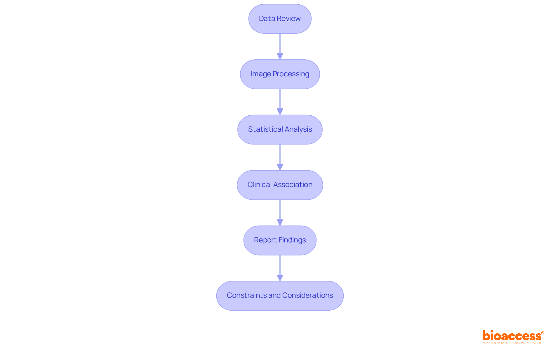
In clinical research, analyzing and interpreting tracer imaging results is essential, involving several key steps that ensure accuracy and relevance.
Data Review: Begin by meticulously examining the gathered visual data to confirm its quality and completeness. This initial step is crucial for identifying any anomalies or gaps that could impact subsequent analyses.
Image Processing: Leverage advanced software tools to process the images, enhancing clarity and enabling quantitative analysis. Contemporary visualization software significantly improves the resolution and detail of the images, facilitating more precise evaluations. For instance, infrared thermography (IRT) has proven effective in diagnosing conditions like diabetic neuropathy and breast cancer, showcasing the practical implications of advanced visual techniques.
Statistical Analysis: Implement appropriate statistical methods to assess the significance of the findings. This includes comparing results against control groups and utilizing techniques such as regression analysis or survival analysis to derive meaningful conclusions from the data. Research indicates that individuals with ≥75% agreement between scan results have a 5-year overall survival rate of 83.3%, underscoring the importance of efficient tracer imaging analysis.
Clinical Association: Link the scanning results to health outcomes by considering individual history and other diagnostic details. This correlation is vital for understanding how scanning results influence patient management and treatment strategies. For example, combining visualization data with patient outcomes has been shown to enhance prognostic evaluations in neuroendocrine tumors.
Report Findings: Compile a comprehensive document detailing the methodology, results, and medical implications of the tracking study. This report should include visual representations of the data, such as graphs or charts, to effectively communicate findings to stakeholders. Incorporating authoritative insights, like the diagnostic significance of infrared temperature measurement, can further bolster the report's credibility.
By adhering to these steps, researchers can effectively analyze and interpret tracer imaging results, providing valuable insights to the field of clinical research. Acknowledging potential constraints, such as sensitivity and specificity concerns in various visualization methods, offers a more balanced perspective on the subject. For instance, the effectiveness of IRT in mass fever screening illustrates the real-world impact of imaging technologies.
Tracer imaging stands as a pivotal advancement in clinical research, leveraging radioactive markers to unveil profound insights into biological processes. This innovative technique not only deepens our understanding of drug distribution and disease progression but also significantly enhances patient outcomes through early diagnosis and personalized treatment strategies.
In this article, we delved into various facets of tracer imaging, emphasizing the importance of advanced visualization methods such as PET and MRI integration. We explored the specific applications of different tracers in oncology and cardiology, providing a comprehensive step-by-step guide for effective implementation. The discussion underscored how these techniques can lead to improved survival rates and more accurate diagnoses, highlighting their essential role in contemporary medical research.
The ongoing evolution of tracer imaging techniques is crucial for propelling personalized medicine and optimizing patient care. As researchers and clinicians adopt these groundbreaking methods, it is vital to prioritize rigorous implementation and analytical practices to fully harness the potential of tracer imaging. By doing so, the medical community can ensure that these advancements translate into meaningful improvements in patient management and treatment outcomes, ultimately reshaping the landscape of clinical research.
What is tracer imaging?
Tracer imaging is a technique that utilizes radioactive materials, known as markers, to observe and measure biological processes within the body.
Why is tracer imaging important in clinical research?
Tracer imaging is crucial in clinical research as it enables the observation of drug distribution, monitoring of disease progression, and assessment of treatment efficacy. It provides real-time insights into physiological functions, enhancing understanding of complex biological systems and ultimately improving patient outcomes.
How does tracer imaging facilitate early diagnosis and treatment decisions?
By providing detailed visualization of biological processes, tracer imaging aids in early diagnosis, informs therapeutic decisions, and supports the development of innovative medical technologies.
What recent advancements have been made in tracer imaging techniques?
Recent advancements include the integration of PET (Positron Emission Tomography) with MRI (Magnetic Resonance Imaging), which has significantly improved diagnostic accuracy, increasing the positive predictive value from 77% to 98%.
What is the role of specialized agents like Ga-FAPI in tracer imaging?
Specialized agents like Ga-FAPI have shown superior tumor absorption and metastatic detection rates compared to traditional methods, particularly in challenging cases such as invasive lobular carcinoma.
How do tracer imaging techniques impact patient outcomes?
Tracer imaging techniques have a substantial impact on patient outcomes, as evidenced by improved survival rates. For example, the five-year survival rate for individuals with HER2-positive breast cancer can reach 91.5% when effectively monitored and treated using advanced scanning methods.
What is the sensitivity of positron emission mammography (PEM) in detecting malignant lesions?
The sensitivity of positron emission mammography (PEM) in identifying malignant lesions has been reported at 96%, highlighting its potential in preoperative staging and treatment planning.
How do survival rates vary among different breast cancer subtypes when monitored with tracer imaging?
The five-year relative survival rates for breast cancer subtypes monitored with tracer imaging show significant variation, with luminal A at 94.4% and luminal B at 90.7%.
What is the future role of tracer imaging in medical research?
As tracer imaging continues to evolve, its role in medical research becomes increasingly vital, paving the way for more personalized and efficient patient care.