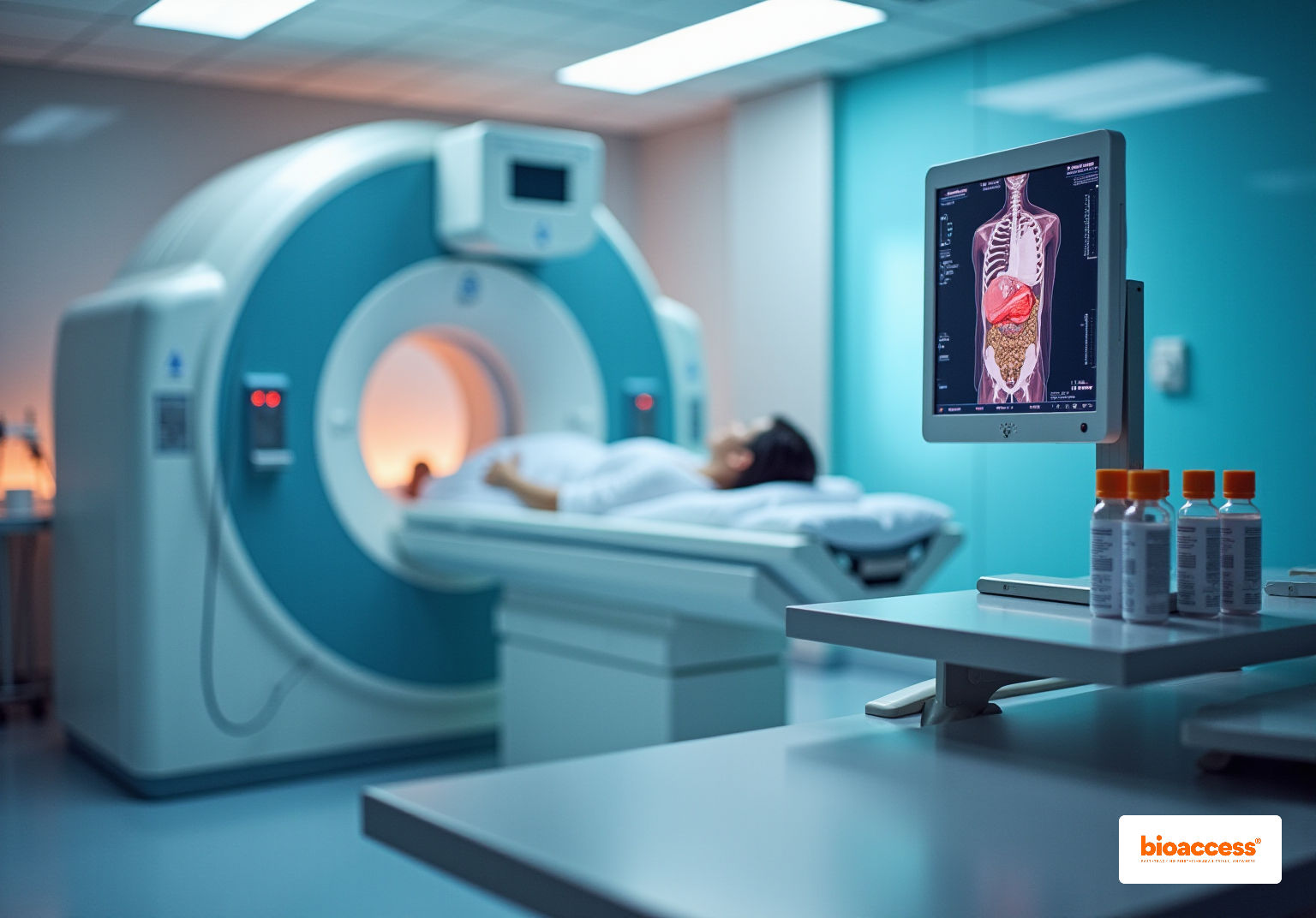


Nuclear medicine is a critical branch of medical imaging that utilizes radiopharmaceuticals to assess the physiological functions of organs and tissues, moving beyond mere structural imaging. This field is underscored by the essential role of radiopharmaceuticals, such as:
These tools are pivotal in nuclear medicine, facilitating early disease detection and enabling targeted treatment strategies, particularly in oncology. The integration of these advanced techniques not only enhances diagnostic accuracy but also significantly improves patient outcomes.
Nuclear medicine stands at the forefront of modern medical imaging, uniquely utilizing radiopharmaceuticals to unveil the physiological workings of the body. This specialized field diverges from traditional imaging techniques by focusing not just on structural anomalies, but also on the functional insights that can significantly impact patient care. As the demand for early disease detection and targeted therapies grows, a pressing question emerges: which branch of medical imaging truly harnesses the power of radiopharmaceuticals to transform diagnostics and treatment in healthcare?
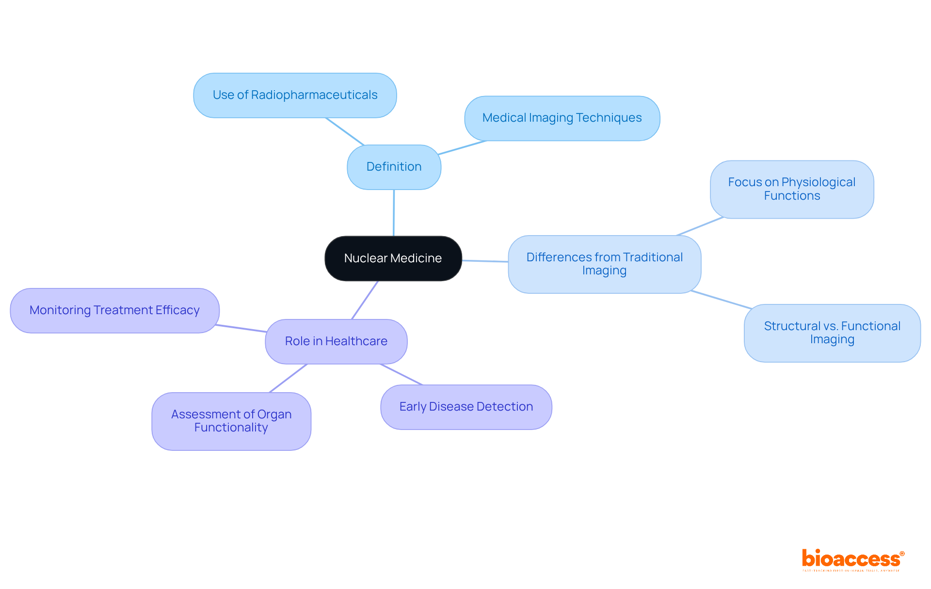
Nuclear therapy is a specialized domain within medical visualization, which is what branch of medical imaging uses radiopharmaceuticals to diagnose and treat a range of illnesses. Unlike traditional imaging techniques such as X-rays or MRIs, which predominantly focus on the structural aspects of the body, nuclear diagnostics prioritizes the physiological functions of organs and tissues. This approach is facilitated by the use of radiopharmaceuticals that emit gamma rays, which raises the question of what branch of medical imaging uses radiopharmaceuticals and can be detected by advanced imaging cameras. The role of nuclear technology in healthcare is indispensable; it allows for the early detection of diseases, assessment of organ functionality, and monitoring of treatment efficacy, thereby enhancing patient care and outcomes.
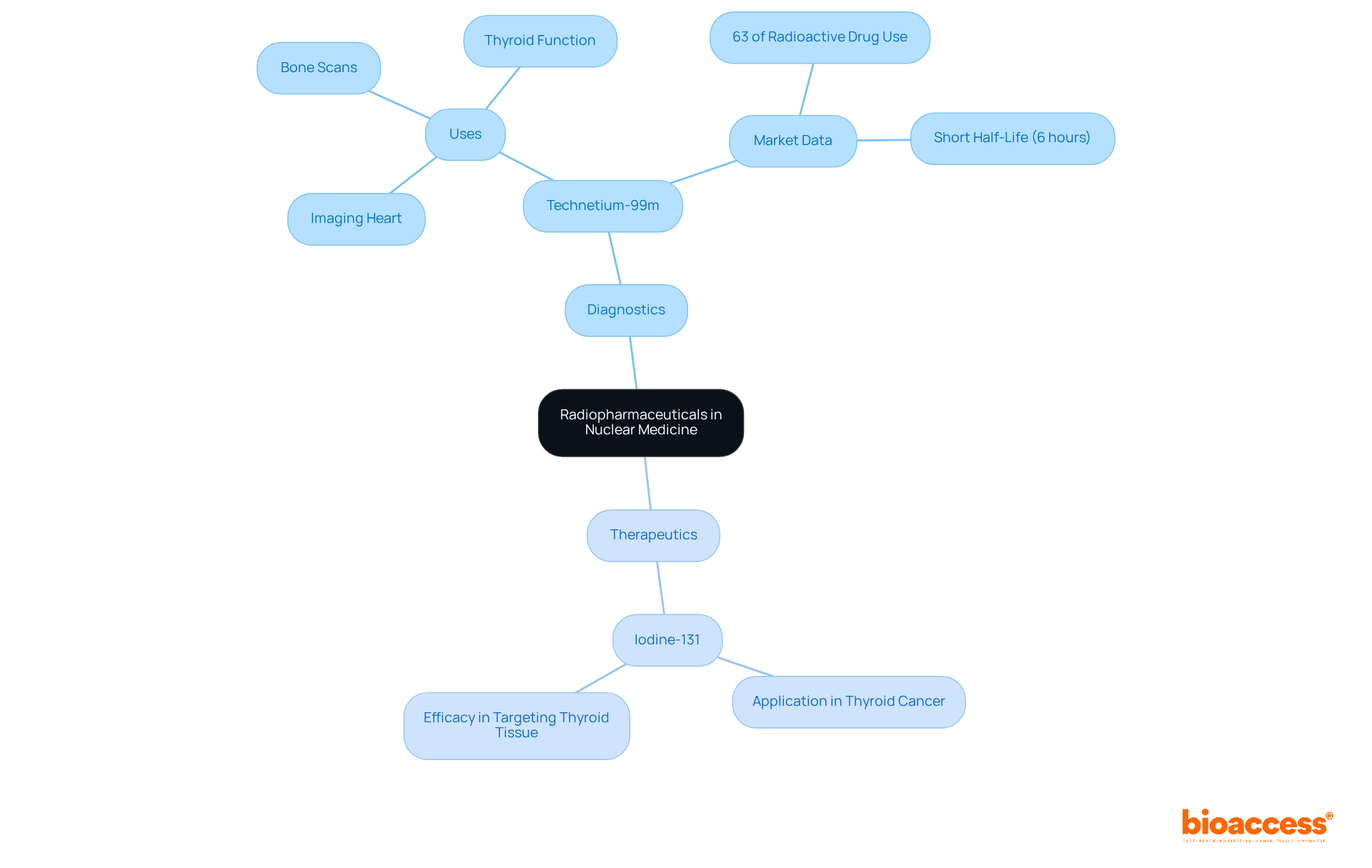
Radiopharmaceuticals, which are specialized compounds containing radioactive isotopes, are crucial in nuclear medicine, specifically in what branch of medical imaging uses radiopharmaceuticals for both diagnostic and therapeutic purposes. These agents can be administered through various routes—such as injection, ingestion, or inhalation—tailored to specific diagnostic or treatment needs.
In diagnostics, what branch of medical imaging uses radiopharmaceuticals to visualize organ function and identify abnormalities, including tumors or infections? For instance, Technetium-99m stands as a cornerstone in modern diagnostics, extensively utilized for imaging the heart, bones, and other organs. Its favorable properties, such as a short half-life of approximately six hours, significantly reduce radiation exposure to patients. Recent market data indicates that Technetium-99m accounts for around 63% of the overall use of radioactive drugs in nuclear medicine, underscoring its critical importance in clinical practice.
In therapeutic contexts, radioactive drugs are meticulously designed to target and eliminate cancer cells effectively. A prominent example is Iodine-131, which has proven successful in treating thyroid cancer, showcasing significant efficacy in targeting thyroid tissue while sparing surrounding healthy cells. Case studies reveal that individuals undergoing Iodine-131 therapy often experience enhanced outcomes, with many achieving remission.
Ongoing research continues to explore what branch of medical imaging uses radiopharmaceuticals, highlighting the efficacy of various radioactive drugs in both diagnostics and therapy and their transformative potential in patient care, particularly in oncology. The adaptability and focused characteristics of these compounds render them indispensable tools in contemporary medical practice, bridging the gap between diagnosis and treatment.
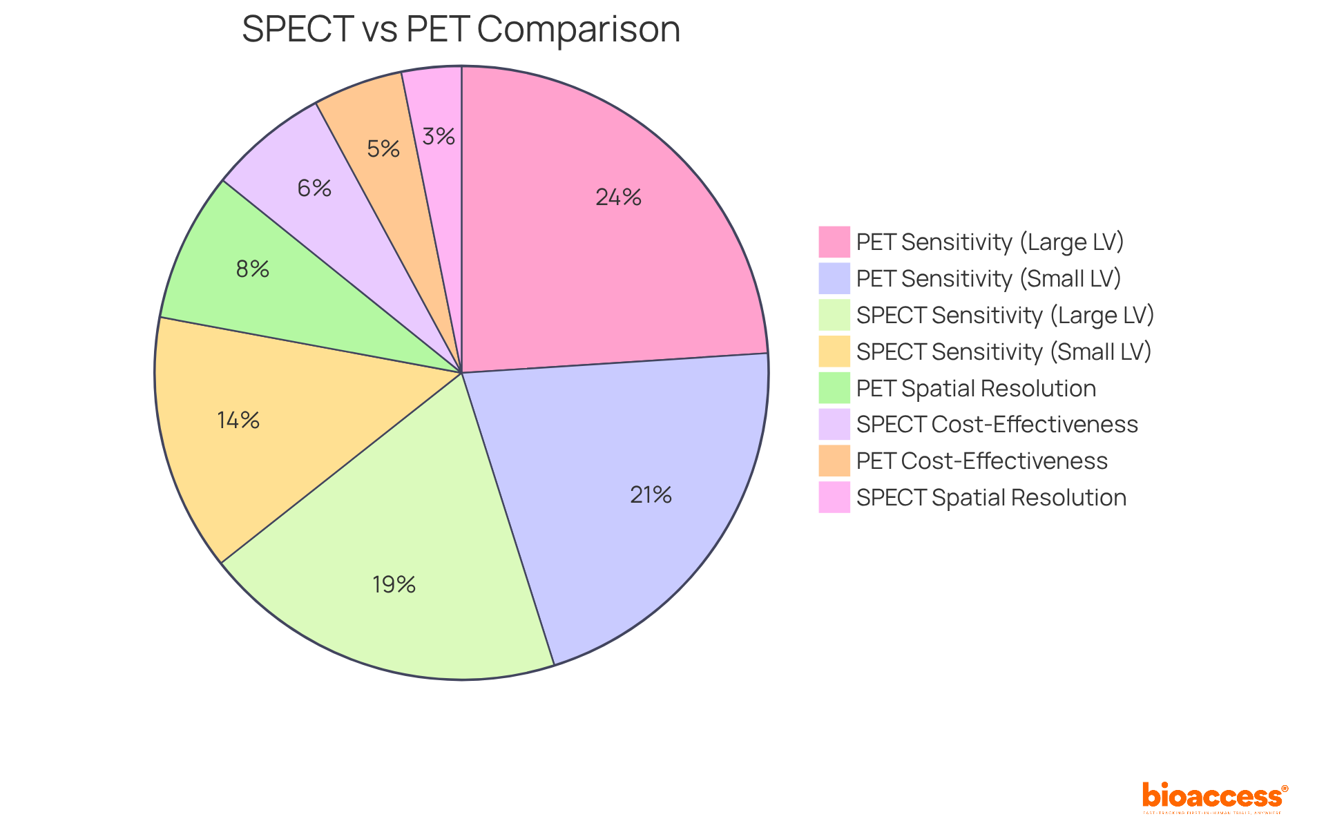
In nuclear medicine, the branch of medical imaging that uses radiopharmaceuticals includes Single Photon Emission Computed Tomography (SPECT) and Positron Emission Tomography (PET), both of which are pivotal diagnostic techniques offering distinct advantages.
Conversely, PET imaging is an example of a branch of medical imaging that uses radiopharmaceuticals, as it identifies positrons emitted by these substances, producing high-resolution images that illuminate metabolic processes. This capability renders PET especially valuable in oncology, where it is utilized to evaluate tumor activity and monitor treatment responses.
Both SPECT and PET are revolutionizing nuclear diagnostics by providing complementary information.
Radiopharmaceuticals are pivotal in the therapeutic landscape of nuclear medicine, particularly in oncology. These agents are meticulously engineered to deliver targeted radiation directly to cancer cells, thereby minimizing damage to surrounding healthy tissues. A prime example is Radium-223, which received FDA approval in 2013 for the treatment of metastatic prostate cancer.
Clinical trials indicate that individuals receiving Radium-223 experience an average life extension of 3.5 months compared to those undergoing standard therapies, with overall survival rates of 14.9 months versus 11.3 months. This targeted approach not only enhances treatment efficacy but also yields improved outcomes for patients. Radium-223 is administered via injection every four weeks for a maximum of six cycles, establishing a structured treatment regimen.
Furthermore, medical isotopes are increasingly utilized in pain management for patients with bone metastases, offering relief and enhancing quality of life. While the therapeutic applications of radioactive drugs underscore their critical role in modern medical imaging, it raises the question of what branch of medical imaging uses radiopharmaceuticals, alongside the challenges such as the shortage of qualified nuclear medicine specialists that remain pertinent.
The transformative potential of these therapies is echoed by experts like Dr. Kunos, who underscores their capacity to revolutionize radiation oncology. Ultimately, the integration of these elements underscores the significance of radiopharmaceuticals in advancing patient care within oncology.

Nuclear medicine emerges as a vital branch of medical imaging, utilizing radiopharmaceuticals to provide unparalleled insights into the physiological functions of organs and tissues. This specialized field distinguishes itself from traditional imaging techniques by emphasizing not only structural abnormalities but also the functional aspects critical for accurate diagnosis and treatment planning. The incorporation of radiopharmaceuticals enhances the ability to detect diseases early, assess organ functionality, and monitor therapeutic responses, significantly elevating patient care.
Key insights reveal the crucial roles of radiopharmaceuticals in both diagnostic and therapeutic contexts. Agents such as Technetium-99m and Iodine-131 exemplify their applications, demonstrating effectiveness in identifying abnormalities and targeting cancer cells, respectively. Furthermore, imaging techniques like SPECT and PET highlight advancements in nuclear diagnostics, offering complementary information that refines the precision of evaluations and treatment strategies. Ongoing research and development in this field underscore the transformative potential of radiopharmaceuticals in modern healthcare.
Embracing advancements in nuclear medicine is essential for healthcare providers and patients alike. With the increasing demand for precise diagnostics and targeted therapies, understanding the significance of radiopharmaceuticals and their applications in medical imaging becomes paramount. Engaging with this knowledge empowers patients to make informed health decisions and encourages healthcare professionals to utilize these innovative tools to optimize patient outcomes. The future of nuclear medicine is poised to be dynamic, with the potential to revolutionize disease diagnosis and treatment, ultimately enhancing the quality of care within healthcare systems worldwide.
What is nuclear medicine?
Nuclear medicine is a specialized field within medical imaging that uses radiopharmaceuticals to diagnose and treat various illnesses, focusing on the physiological functions of organs and tissues.
How does nuclear medicine differ from traditional imaging techniques?
Unlike traditional imaging techniques such as X-rays or MRIs, which primarily focus on the structural aspects of the body, nuclear medicine emphasizes the physiological functions of organs and tissues.
What are radiopharmaceuticals?
Radiopharmaceuticals are substances that emit gamma rays and are used in nuclear medicine to facilitate the diagnosis and treatment of diseases.
What are the key roles of nuclear medicine in healthcare?
Nuclear medicine plays a crucial role in early disease detection, assessment of organ functionality, and monitoring of treatment efficacy, ultimately enhancing patient care and outcomes.
What type of imaging is used to detect radiopharmaceuticals?
Advanced imaging cameras are used to detect the gamma rays emitted by radiopharmaceuticals in nuclear medicine.