


This article delves into ten essential types of nuclear medicine and their applications, underscoring the diverse roles these techniques play in diagnosing and treating a range of medical conditions. By detailing specific imaging methods such as PET and SPECT, it highlights how these technologies enhance diagnostic accuracy and treatment efficacy, particularly in fields like oncology, cardiology, and neurology. This showcases their critical importance in modern healthcare, emphasizing the need for continued collaboration and innovation in clinical research.
In the ever-evolving Medtech landscape, nuclear medicine stands out as a pivotal player in addressing key challenges. The integration of advanced imaging techniques not only improves patient outcomes but also streamlines the diagnostic process, making it vital for healthcare professionals to stay informed about these developments. As we explore the impact of these methods, consider how they might address your own challenges in clinical research.
Ultimately, the significance of nuclear medicine in enhancing healthcare cannot be overstated. By fostering collaboration among researchers, clinicians, and technologists, we can pave the way for groundbreaking advancements that will shape the future of medical diagnostics and treatment.
Nuclear medicine is at the cutting edge of modern healthcare, providing innovative diagnostic and therapeutic solutions that utilize radioactive materials to visualize and treat a wide range of diseases. As this field evolves, it becomes increasingly important for healthcare professionals and patients to grasp the various types of nuclear medicine and their applications.
What challenges and advancements are shaping the future of nuclear medicine? How can these technologies enhance patient outcomes?
This exploration delves into key techniques, emerging trends, and the essential role of collaboration in advancing nuclear medicine practices.
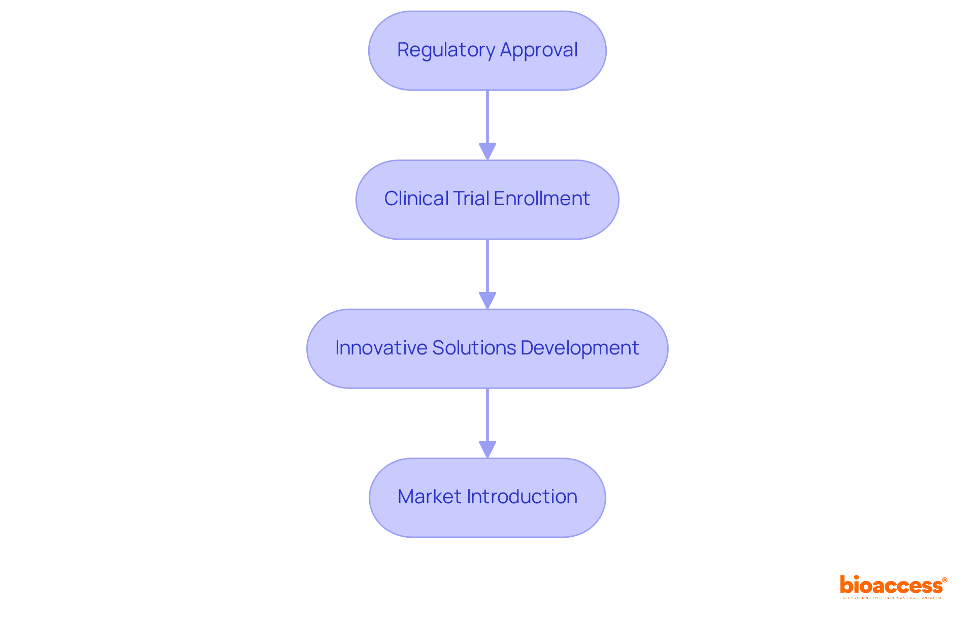
bioaccess® plays a pivotal role in advancing radiological research across Latin America, leveraging the region's regulatory efficiency and diverse consumer groups. With ethical approvals secured in just 4-6 weeks, bioaccess® accelerates enrollment in clinical trials, significantly reducing the time needed to bring innovative radiological solutions to market. This agility is essential for Medtech and Biopharma firms eager to capitalize on emerging opportunities in various nuclear medicine types, ensuring that groundbreaking therapies reach patients faster than traditional markets allow.
In the rapidly evolving Medtech landscape, bioaccess® addresses key challenges faced by companies striving to innovate. The ability to navigate regulatory processes swiftly not only enhances operational efficiency but also fosters a competitive edge. As firms seek to introduce novel treatments, the collaboration with bioaccess® becomes increasingly valuable, enabling them to respond to market demands with agility and precision.
Ultimately, the importance of collaboration in clinical research cannot be overstated. By partnering with bioaccess®, organizations can streamline their processes and enhance their capacity to deliver cutting-edge therapies. As the landscape continues to evolve, taking proactive steps now will position firms for success in the future.
Nuclear therapy is one of the nuclear medicine types, standing as a specialized field that employs radioactive materials for diagnosing and treating a diverse range of diseases. Its relevance in contemporary healthcare cannot be overstated, as it provides unique insights into the physiological functions of organs and tissues. By utilizing radiopharmaceuticals, healthcare professionals can effectively visualize and assess conditions such as cancer, cardiovascular diseases, and neurological disorders. For instance, myocardial perfusion imaging demonstrates a diagnostic sensitivity of 80% and specificity of 92% for coronary artery disease, showcasing how this field enhances diagnostic accuracy.
Moreover, the ability to detect illnesses at an early stage significantly improves treatment outcomes, underscoring the vital role of various nuclear medicine types in patient care. Recent advancements and ongoing research in various nuclear medicine types are broadening its applications, particularly in cancer treatment, where targeted therapies are increasingly integrated into clinical practice. In-pentetreotide scintigraphy, with a sensitivity of approximately 85% for carcinoid assessment, exemplifies current trends in this field.
Furthermore, with around 40 million diagnostic radiological procedures performed annually, the importance of nuclear therapy is evident. As the field evolves, the emphasis on radiation safety and effective communication regarding the benefits and risks of nuclear health practices remains paramount. This is especially crucial given the involvement of eight international organizations in discussions on radiation protection, ensuring that both patients and healthcare providers are well-informed.

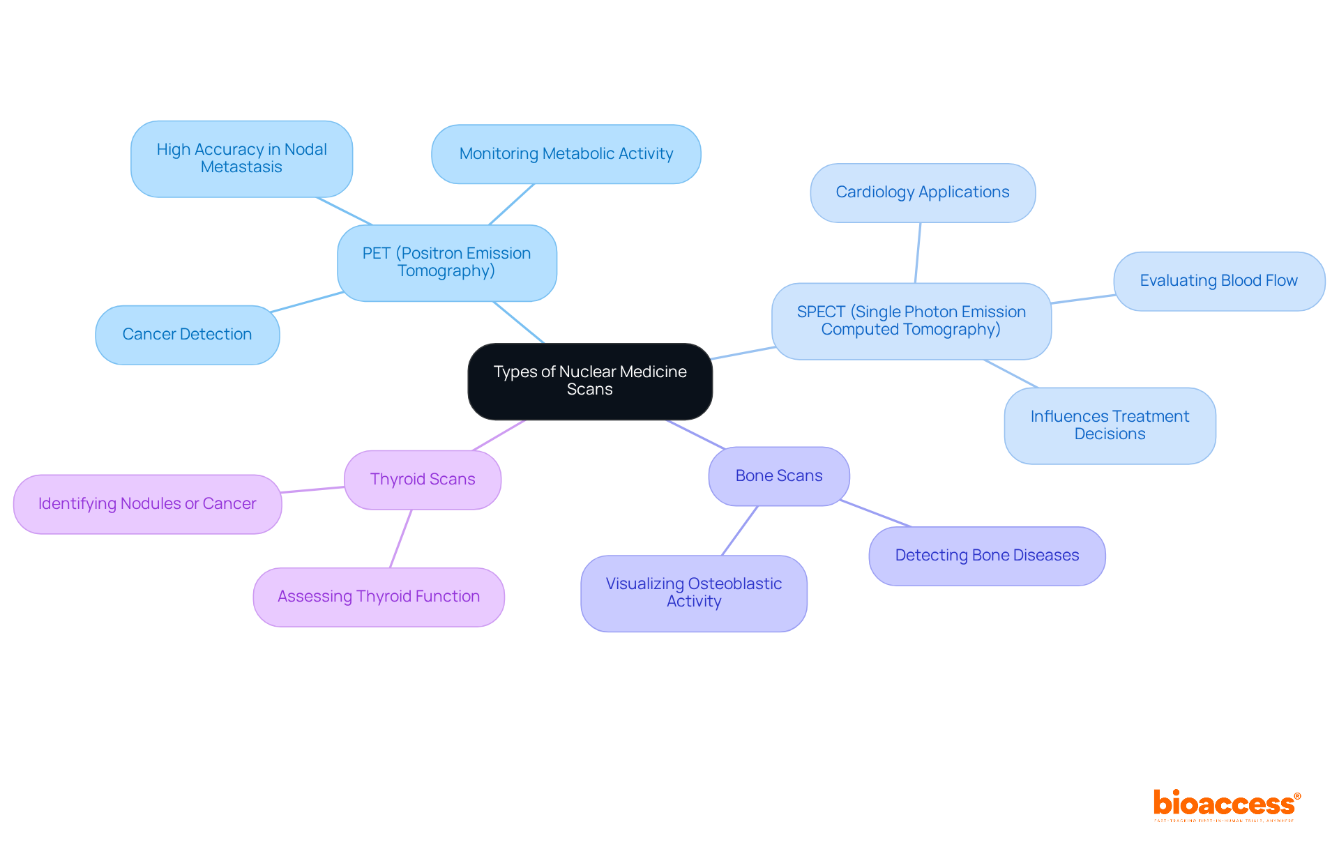
Nuclear medicine plays a pivotal role in modern diagnostics, encompassing several key scanning techniques that provide unique insights into various medical conditions:
PET (Positron Emission Tomography): This technique is primarily utilized for cancer detection and monitoring. PET scans deliver detailed images of metabolic activity, making them invaluable in identifying malignancies. Recent studies indicate that FDG-PET demonstrates significantly higher accuracy in detecting distant nodal metastasis compared to conventional techniques, boasting a negative predictive value of 91% versus 84% for CT in detecting N2 lymph nodes. This underscores its critical role in oncology.
SPECT (Single Photon Emission Computed Tomography): Widely used in cardiology, SPECT scans evaluate blood flow and heart function. They are essential for diagnosing conditions such as coronary artery disease and assessing myocardial viability. The integration of SPECT with CT scans enhances diagnostic capabilities, with studies indicating that this combination can influence treatment decisions by 25% to 30%.
Bone Scans: These scans are instrumental in detecting bone diseases, including fractures and infections. Utilizing radiopharmaceuticals, they visualize areas of increased osteoblastic activity, providing insights into conditions like metastatic bone disease.
Thyroid Scans: Employed to assess thyroid function, these scans help identify abnormalities such as nodules or cancer. They play a crucial role in managing thyroid disorders, guiding treatment decisions based on functional scan results.
Each of the nuclear medicine types is essential in the diagnostic toolkit, enhancing outcomes for individuals through accurate visualization and targeted treatments. As PET scanning continues to gain recognition for its emerging applications across various medical fields, it remains a valuable radiological tool. Proper patient preparation, such as avoiding certain foods or medications before scans, is also crucial for achieving optimal results.
Radiotheranostics represents a groundbreaking advancement in nuclear medicine, seamlessly integrating diagnostic imaging with targeted therapy. This innovative approach allows for the precise delivery of therapeutic agents directly to cancer cells, all while monitoring treatment effectiveness through imaging.
For instance, the application of radiolabeled antibodies not only assists in tumor identification but also enhances treatment efficacy, significantly minimizing harm to surrounding healthy tissues. This dual capability not only improves treatment outcomes, as demonstrated by enhanced patient responses in clinical trials, but also personalizes patient care, marking a significant leap forward in oncology.

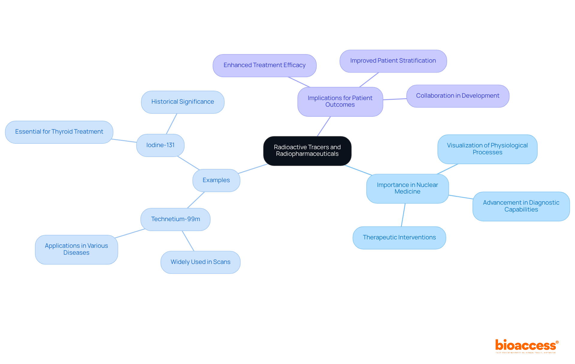
Radioactive tracers and radiopharmaceuticals are vital components of various nuclear medicine types, enabling the visualization and treatment of different illnesses. These substances emit detectable radiation, allowing clinicians to observe physiological processes in real-time. For instance, Technetium-99m is widely used in numerous scans, while Iodine-131 is essential for treating thyroid conditions. The development and application of these tracers are not just beneficial; they are vital for advancing the diagnostic capabilities and therapeutic interventions associated with nuclear medicine types.
As we delve deeper into the Medtech landscape, it's clear that the integration of these technologies addresses key challenges in clinical research. The ability to visualize and treat conditions effectively can significantly enhance patient outcomes. How can we leverage these advancements to tackle the pressing issues in healthcare today?
In summary, the importance of collaboration in the development of radioactive tracers cannot be overstated. Continued innovation in nuclear medicine types is essential for enhancing diagnostic and therapeutic practices, paving the way for a healthier future.
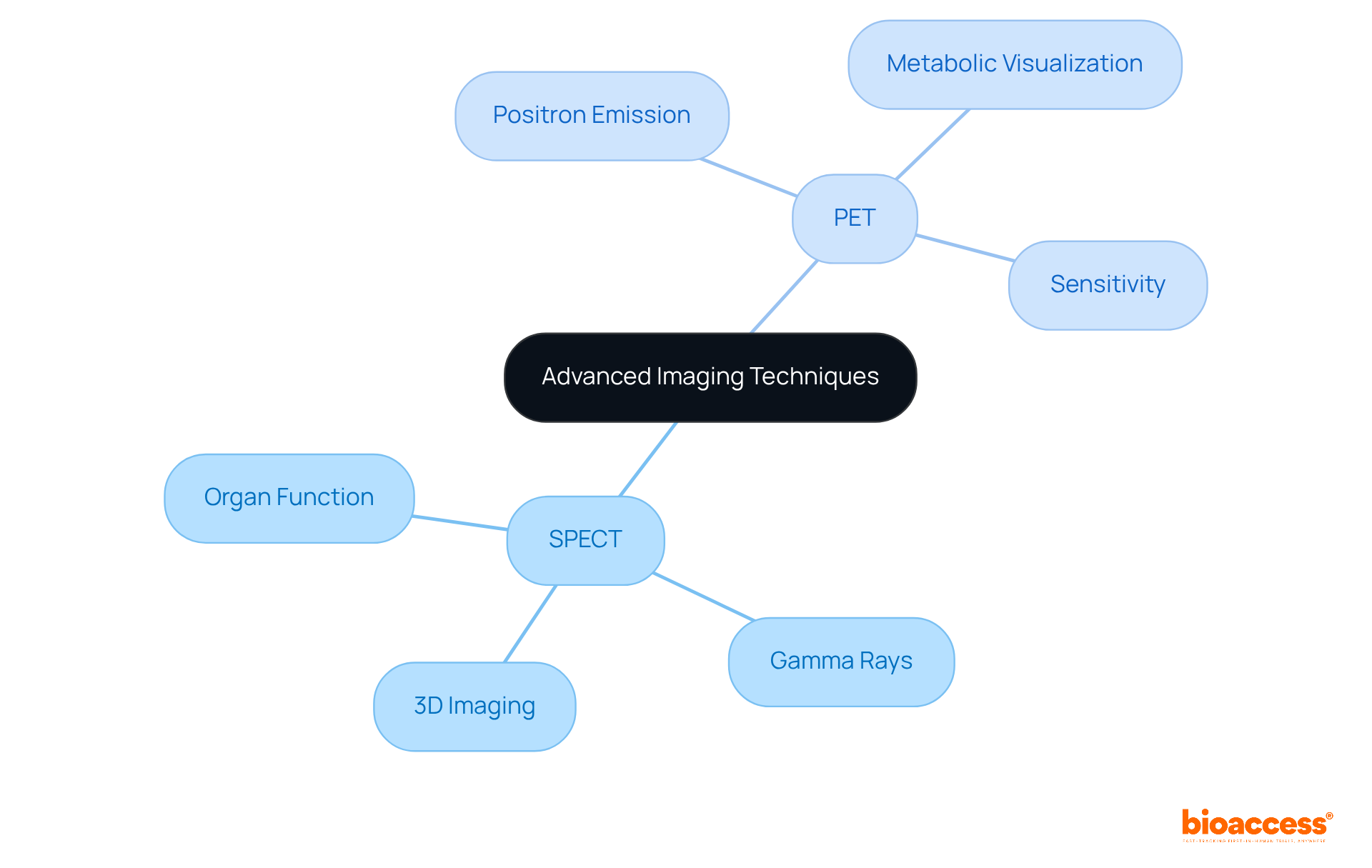
SPECT and PET are advanced scanning techniques that have significantly transformed nuclear medicine. These modalities not only provide critical insights into organ function and blood flow but also play a pivotal role in diagnosing various conditions. SPECT delivers three-dimensional images by detecting gamma rays emitted from radiotracers, enabling detailed assessments. In contrast, PET utilizes positron-emitting radiotracers, offering superior sensitivity and specificity to visualize metabolic processes. This enhanced capability is crucial for diagnosing a range of conditions, including cancer, heart disease, and neurological disorders.
For instance, PET scans demonstrate a remarkable sensitivity of 82.5% for identifying metastasis, compared to just 53.6% for CT scans. This stark contrast underscores the significance of these diagnostic techniques in clinical decision-making. Notably, PET also shows an improved sensitivity of +11.5% for nodal involvement compared to CT, further emphasizing its diagnostic advantages. The median age reported in US cancer statistics is 66 years, providing context for the demographics affected by these conditions.
Recent advancements in visual technology, such as iterative reconstruction techniques, have further enhanced picture quality and diagnostic precision. This solidifies the role of SPECT and PET in modern healthcare. As Daniel Steinemann noted, the increased sensitivity of PET/CT allows for improved detection of both lymph node metastasis and distant metastasis, reinforcing the value of these imaging modalities in clinical practice. The integration of these technologies into routine diagnostics not only improves patient outcomes but also enhances the overall efficacy of clinical research.
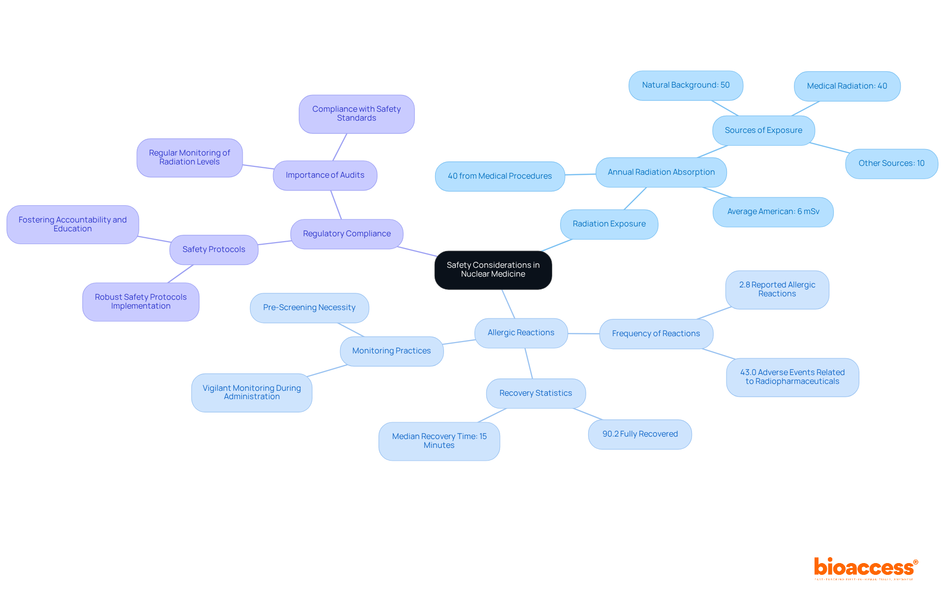
While radiological therapy offers significant advantages, understanding the associated risks is crucial for patient safety. Key safety considerations include:
Radiation Exposure: Patients typically receive low levels of radiation during nuclear medicine scans, which are generally considered safe. However, monitoring exposure levels is vital, as approximately 40% of the average American's radiation exposure comes from medical procedures, including diagnostic imaging and various nuclear medicine types. On average, the typical American absorbs about 6 mSv of radiation annually, with substantial contributions from both natural and medical sources.
Allergic Reactions: Allergic responses to radiopharmaceuticals can occur, with a reported frequency of 2.8% among individuals. Furthermore, 43.0% of patient-reported adverse events were possibly or probably related to radiopharmaceuticals. This highlights the necessity for thorough pre-screening and vigilant monitoring during and after administration to effectively manage potential adverse events. Notably, 90.2% of individuals who reported adverse events made a complete recovery, with a median recovery duration of just 15 minutes.
Regulatory Compliance: Strict adherence to regulatory guidelines is essential for ensuring that all radioactive treatment procedures are conducted safely and ethically. Regular audits and monitoring of radiation exposure levels help maintain compliance with safety standards, fostering a culture of accountability and education among healthcare providers. By implementing robust safety protocols, healthcare professionals can significantly reduce risks and enhance patient trust in radiological practices.
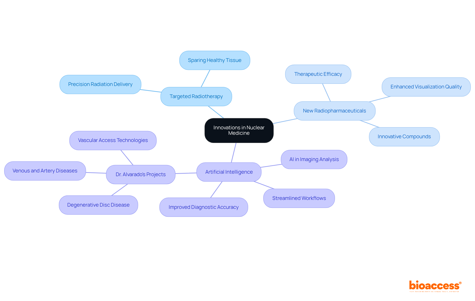
Innovations in various nuclear medicine types are rapidly evolving, propelled by significant advancements in technology and research. Current areas of focus include:
Dr. Sergio Alvarado, a Clinical Trial Manager at bioaccess, underscores the importance of innovative medical solutions in Latin America, where access to advanced research can significantly improve health outcomes. With a robust background in medical affairs at Novartis and experience as a general practitioner at Colsubsudio, he is exceptionally equipped to spearhead advancements in this field. His initiatives in artificial intelligence aim to identify medical issues more efficiently, showcasing how these technologies can revolutionize medical practices. Currently, he is engaged in projects addressing degenerative disc disease, venous and artery diseases, and vascular access technologies. These innovations promise to enhance the capabilities of nuclear healthcare by utilizing different nuclear medicine types, ultimately improving patient outcomes and expanding treatment options.
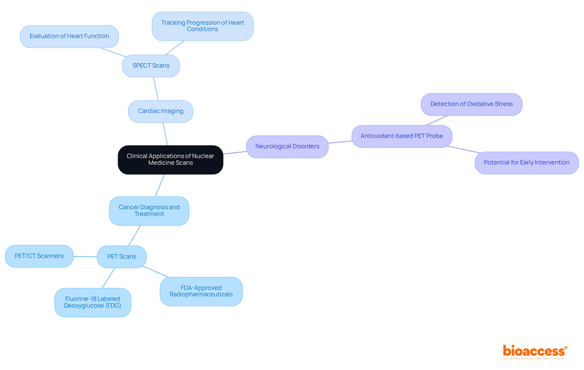
Nuclear medicine types of scans play a crucial role in modern healthcare, offering a range of clinical applications that are vital for patient care.
Cancer Diagnosis and Treatment: Positron Emission Tomography (PET) scans are indispensable for detecting and monitoring various cancers. The use of fluorine-18 labeled deoxyglucose (FDG) stands out as the most effective radiopharmaceutical for this purpose. PET/CT scanners have emerged as the primary imaging tool for cancer staging globally. Recent advancements have led to FDA-approved radiopharmaceuticals that aid in the precise diagnosis of conditions like Alzheimer’s disease, underscoring the evolving significance of nuclear therapy in oncology.
Cardiac Imaging: Single Photon Emission Computed Tomography (SPECT) scans are essential for evaluating heart function and blood flow, particularly in diagnosing coronary artery disease. These scans provide three-dimensional images that are instrumental in tracking the progression of heart conditions, making them a critical asset in cardiology.
Neurological Disorders: The application of nuclear medicine is expanding in the diagnosis of neurological conditions such as Alzheimer's and Parkinson's diseases. Innovative imaging techniques, including a recent antioxidant-based PET probe developed by researchers at St. Jude Children’s Research Hospital and the University of Virginia, enable the detection of oxidative stress in neurodegeneration, highlighting the potential for early intervention.
While radioactive scans are generally safe, some individuals may experience complications such as pain or swelling at the injection site, with rare reactions including fever or allergic responses. These applications illustrate the versatility and significance of nuclear medicine types in diagnosing and treating a wide array of illnesses, reinforcing their essential role in enhancing patient care.
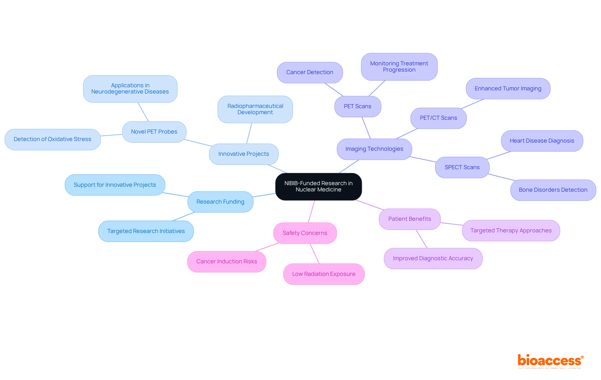
The National Institute of Biomedical Imaging and Bioengineering (NIBIB) plays a crucial role in advancing radiological science through targeted research funding. By supporting innovative projects, NIBIB not only fosters the development of new imaging technologies and radiopharmaceuticals but also enhances treatment methodologies. This funding deepens our scientific understanding of radiology and translates into improved clinical practices, ultimately benefiting patients.
Current NIBIB-supported research projects are paving the way for the next generation of radiological applications. For instance, researchers at St. Jude Children’s Research Hospital and the University of Virginia have developed novel PET probes designed to detect oxidative stress, a critical factor for early interventions in neurodegenerative diseases. The primary purpose of PET scans is to identify cancer and monitor its progression, underscoring the significance of these advancements in enhancing diagnostic accuracy and treatment efficacy.
While the benefits of these imaging technologies are substantial, it is essential to address concerns regarding low radiation exposure in medical imaging. Although this exposure is considered minimal compared to the benefits, it remains a relevant aspect of advancements in nuclear medicine types. As we continue to explore these technologies, collaboration and awareness will be key in navigating the challenges and opportunities that lie ahead.
The exploration of nuclear medicine underscores its vital role in modern healthcare, showcasing a range of techniques and applications that enhance both diagnostic and therapeutic practices. This field not only offers unique insights into diseases but also enables targeted treatments that significantly improve patient outcomes. The integration of advanced imaging technologies, such as PET and SPECT, highlights the remarkable accuracy of nuclear medicine in detecting conditions like cancer and cardiovascular diseases.
Key advancements in radiopharmaceuticals and innovative approaches like radiotheranostics, which merge diagnosis and treatment, are pivotal. Ongoing research funded by organizations such as NIBIB propels the field forward, paving the way for new imaging technologies and therapeutic strategies. With safety considerations remaining paramount, a commitment to regulatory compliance and patient education ensures that the benefits of nuclear medicine can be harnessed effectively while minimizing risks.
In light of these insights, the continued evolution of nuclear medicine is essential for addressing today’s pressing healthcare challenges. Emphasizing collaboration, innovation, and patient-centered approaches will not only enhance diagnostic accuracy but also expand treatment options, ultimately leading to a healthier future. Engaging with these advancements and supporting ongoing research initiatives will be crucial in maximizing the potential of nuclear medicine to improve patient care across diverse medical landscapes.
What is bioaccess® and how does it contribute to nuclear medicine research in Latin America?
bioaccess® plays a crucial role in advancing radiological research in Latin America by leveraging the region's regulatory efficiency and diverse consumer groups. It accelerates enrollment in clinical trials, securing ethical approvals in just 4-6 weeks, which helps bring innovative radiological solutions to market faster.
Why is the speed of regulatory processes important for Medtech and Biopharma firms?
The ability to navigate regulatory processes swiftly enhances operational efficiency and provides a competitive edge for Medtech and Biopharma firms. This agility allows them to respond to market demands and introduce novel treatments more effectively.
How does collaboration with bioaccess® benefit organizations in clinical research?
Partnering with bioaccess® streamlines processes and enhances the capacity of organizations to deliver cutting-edge therapies. This collaboration is increasingly valuable as the landscape of clinical research evolves.
What is nuclear medicine and why is it important?
Nuclear medicine is a specialized field that uses radioactive materials for diagnosing and treating various diseases. It provides unique insights into the physiological functions of organs and tissues, significantly improving diagnostic accuracy and treatment outcomes, particularly in conditions like cancer and cardiovascular diseases.
What are some key techniques used in nuclear medicine scans?
Key techniques include:
How do PET and SPECT scans differ in their applications?
PET scans are primarily utilized for cancer detection and monitoring metabolic activity, while SPECT scans are widely used in cardiology to evaluate blood flow and heart function. Both techniques enhance diagnostic capabilities when integrated with other imaging methods.
What role do radiopharmaceuticals play in nuclear medicine?
Radiopharmaceuticals are used in various nuclear medicine scans to visualize and assess conditions, providing insights into diseases such as cancer, cardiovascular issues, and bone disorders.
What is the significance of radiation safety in nuclear medicine?
Radiation safety is paramount in nuclear medicine due to the involvement of radioactive materials. Effective communication regarding the benefits and risks of nuclear health practices ensures that both patients and healthcare providers are well-informed, supported by discussions among international organizations on radiation protection.